
SAMSUN, Türkiye – In a new case report, researchers Dr. Güler Silov from Samsun University Faculty of Medicine and Dr. Aslı Ayan from Gulhane Training and Research Hospital shed light on a significant, albeit rare, side effect of long-term warfarin therapy: extensive tracheobronchial cartilage calcification. Their study, published in the inaugural issue of The Journal of Diagnosis and Therapy in Imaging, uniquely utilizes both pre- and post-treatment hybrid imaging to document the development of this condition over a seven-year period.
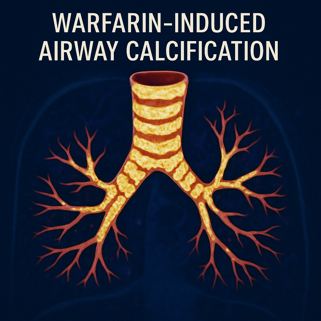
The case centers on a 74-year-old female patient who was undergoing a Positron Emission Tomography/Computed Tomography (PET/CT) scan for the metabolic evaluation of newly identified pulmonary nodules. While the PET/CT scan confirmed the nodules were not metabolically active—showing no abnormal Fluorodeoxyglucose (18F-FDG) uptake—it made a serendipitous discovery: diffuse and striking calcification throughout the patient’s tracheal and bronchial cartilage. Importantly, this extensive calcification was also found to be metabolically inert.
To understand the timeline of this finding, the clinical team reviewed the patient’s archived medical images. A Technetium-99m Macroaggregated Albumin (99mTc-MAA) Single Photon Emission Computed Tomography/Computed Tomography (SPECT/CT) scan, performed seven years prior to diagnose a pulmonary embolism, provided a crucial baseline. The CT component of that earlier scan showed no evidence of the irregular or nodular calcifications that were now prominent.
The patient had been on continuous, long-term warfarin sodium therapy for atrial fibrillation and since the diagnosis of the pulmonary embolism seven years ago. Laboratory tests ruled out other potential causes, such as hypercalcemia or hyperphosphatemia.
While some degree of tracheobronchial calcification can be a normal physiological process associated with aging, particularly in women, the extensive nature seen in this patient pointed towards a different etiology. By comparing the pristine condition of the airways on the initial SPECT/CT with the widespread calcification on the recent PET/CT, the authors compellingly illustrate the calcification’s development during the period of anticoagulation therapy.
This report serves as an important clinical reminder that long-term warfarin use can be a causative factor in profound tracheobronchial calcification. The authors effectively demonstrate the diagnostic power of hybrid imaging modalities like SPECT/CT and PET/CT in not only identifying primary pathologies but also in monitoring and uncovering long-term, treatment-induced changes.
This report highlights:
- The potential for long-term warfarin sodium therapy to induce significant, diffuse calcification of the tracheobronchial cartilage.
- The value of comparing sequential hybrid imaging scans (SPECT/CT and PET/CT) over several years to document the progression of such pathological changes.
- The characteristic finding that this type of warfarin-induced calcification is metabolically inert, showing no abnormal 18F-FDG uptake on PET/CT.
- The importance of considering medication history, specifically anticoagulants like warfarin, in the differential diagnosis of extensive airway calcification.
To cite this article: Silov G, Ayan A. Pre- and post-treatment hybrid imaging findings in a case of tracheobronchial cartilage calcification induced by long-term warfarin sodium therapy. TheJODTi. 2025;1(1):20-22.