
The Journal of Diagnostic and Theranostic Imaging
Volume 1, Issue 1, Pages 20-22 (June 2025)
Received: 31 May 2025 • Accepted: 19 June 2025 • Published: 29 June 2025
Letter to the Editor
Pre- and Post-treatment Hybrid Imaging Findings in a Case Tracheobronchial Cartilage Calcification Induced by Long-term Warfarin Sodium Therapy
Güler Silov¹, Aslı Ayan²
¹Department of Nuclear Medicine, Samsun University Faculty of Medicine, Samsun, Türkiye.
²Department of Nuclear Medicine, University of Health Sciences, Gulhane Training and Research Hospital, Ankara, Türkiye.
To the Editor,
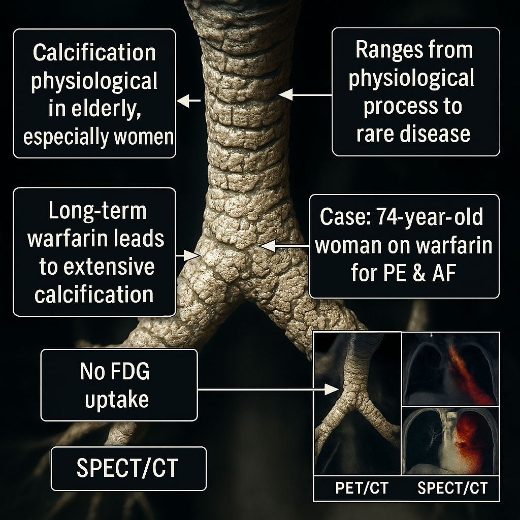
Calcification of the tracheobronchial cartilage is considered a normal physiological process in older patients, especially in women. It ranges from a physiological process to a rare manifestation of certain diseases with extensive calcifications. Long-term warfarin sodium treatment may also lead to extensive tracheobronchial calcification. Here we report a case of diffuse calcification of the tracheobronchial cartilage as a result of long-term anticoagulant therapy in a patient on warfarin sodium therapy for pulmonary embolism and atrial fibrillation, with pretreatment SPECT/CT and post-treatment PET/CT hybrid imaging findings.
A 74-years old woman underwent PET/CT for the metabolic characterization of newly appearing pulmonary nodules. PET/CT revealed no increased FDG uptake in the pleural-based lesion (approximately 17×16 mm) and millimetric nonspecific nodules in the right middle lobe. In addition, no pathological FDG uptake was detected in the air cystic structures and millimetric peripheral calcification was accompanied by fibrotic changes in the superior segment of the right lower lobe.
Diffuse calcification of the tracheobronchial tree without abnormal FDG uptake was observed (Figure 1). The patient’s laboratory test results were neither hypercalcemic nor hyperphosphatemic. Archived files were searched to compare the findings. The seven years old Tc-99m MAA SPECT/CT image showed a segmental perfusion defect in the normal parenchyma with a reported diagnosis of pulmonary embolism, without any evidence of nodular or irregular tracheal or bronchial calcification at the time of diagnosis (Figure 2).
Share this:
- Share on Facebook (Opens in new window) Facebook
- Share on X (Opens in new window) X
- Share on LinkedIn (Opens in new window) LinkedIn
- Share on WhatsApp (Opens in new window) WhatsApp
- Share on Telegram (Opens in new window) Telegram
- Share on Reddit (Opens in new window) Reddit
- Share on Threads (Opens in new window) Threads
- Email a link to a friend (Opens in new window) Email
- Print (Opens in new window) Print